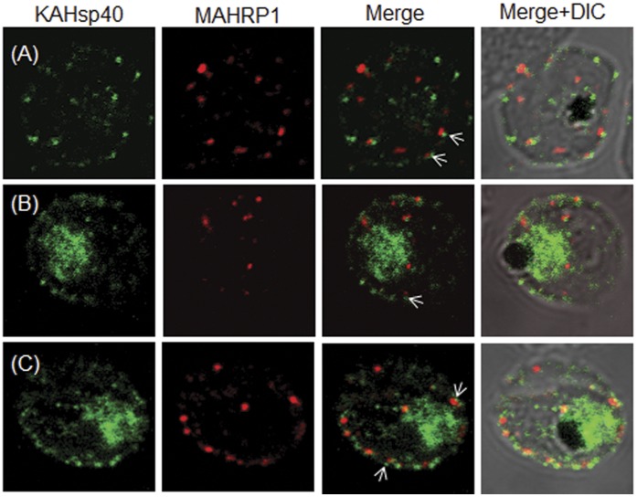
Figure 3. KAHsp40 does not associate with Maurer’s cleft.
(A–C) IFA reveals that both KAHsp40 and MAHRP1 are present in discrete foci in the infected erythrocyte, however, they do not co-localize with each other in spite of signals being in close proximity (highlighted by white arrows). The images shown have been taken at the trophozoite stage.

