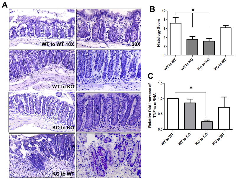
Figure 2. Parameters of DSS-induced colitis are attenuated in the absence of mucosal- A2BAR.
H&E staining was performed on colonic sections from bone marrow transplant recipients and visualized at 10X and 20X (A). Histological scores were blindly calculated and averaged for 5–6 mice/group, where the error bars represent standard deviation. The histological scores were based on the following criteria: Inflammatory cells in the laminia propria (rare inflammatory cells = 0, increased number of granulocytes = 1, confluence of inflammatory cells = 2, transmural extension of infiltrate = 3), crypt damage (intact crypt = 0, loss of 1/3 of the basal crypt = 1, loss of 2/3 of the basal crypt = 2, entire crypt loss = 3, change of epithelial surface with erosion = 4, confluent erosion = 5) and ulcer evaluation (no ulcer = 0, 1–2 foci of ulcers = 1, 3–4 foci = 2, extensive ulceration = 3). The total score of these criteria gave a histological score ranging from 0 to 11 (B). TNF-α mRNA levels were measured from isolated colonic RNA using real-time PCR. 36B4 was used as an internal control to account for variances between samples (C). Statistical significance was assessed using Students t-test, where *, p < .05 and the error bars represent the calculated standard deviations.

