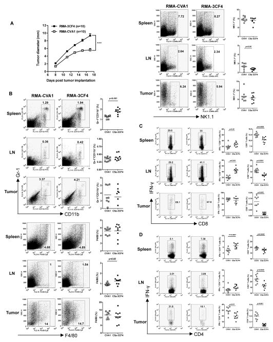
Figure 5. C5a-expressing lymphoma cells have significantly enhanced tumor progression.
(A) Wildtype C57Bl/6 mice were injected with C5a-expressing lymphoma RMA-3CF4 cells or RMA CVA1 cells (n=10) and tumor growth was recorded. Data are shown as mean±s.e.m. ***p<0.001. (B) Spleen, TDLN, and tumor from tumor-bearing mice were prepared for single cell suspensions. Cells were then stained with Gr-1, CD11b, F4/80, or NK1.1. Representative dot plots and summarized data are shown. (C) Cells were stimulated with PMA/ionomycin and surface stained with CD8 and IFN-γ intracellularly. Representative dot plots (cells were gated on the CD8+ T cells), summarized IFN-γ-producing CD8+ T cells, and total CD8+ T cells are shown. (D) Cells were stimulated with PMA/ionomycin and surface stained with CD4 and IFN-γ intracellularly. Representative dot plots (cells were gated on the CD4+ T cells), summarized IFN-γ-producing CD4+ T cells, and total CD4+ T cells are shown.

