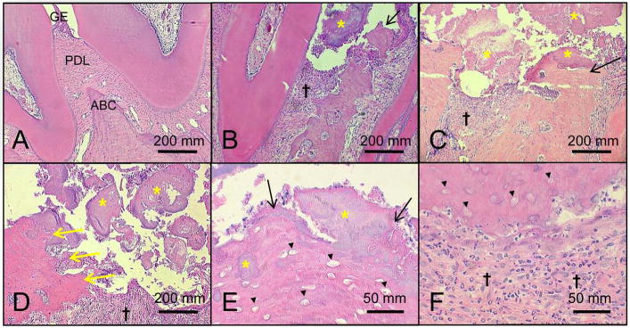
Figure 2. ONJ-like lesions develop in mandibles of rice rats that received a high dose (HD, oncologic doses) of zoledronic acid (ZOL) for 18 wks.

Representative histologic photos of mandibles from female rice rats treated with vehicle (A) and HD-ZOL for 18 wks (B-F). Photos were taken at the interproximal space between the first and second molars. Note the normal histologic features of the gingival epithelium (GE), periodontal ligament (PDL) and the alveolar bone crest (ABC) in a mandible from a rice rat treated with vehicle for 18 wks (A). In contrast, ulceration of the gingival epithelium accompanied by exposed necrotic alveolar bone (black arrows) (B, C and E), osteolysis (yellow arrows) (D), moderate to severe inflammatory cell infiltration (†) (B-D and F), and massive accumulation of bacterial plaque (yellow asterisk) (B-E) are frequently observed in mandibles of rice rats treated with HD-ZOL. Note bacterial plaque on the surface of the ulcerated epithelium, attached to the ABC surface, or embedded in the necrotic bone (yellow asterisk) (B-E). The osteolysis in the HD-ZOL rats (yellow arrows) was sometimes so severe that all the alveolar bone and part of the body of the mandible disappeared (yellow arrows) (D). Necrotic alveolar bone was histologically characterized by extensive areas of tissue with empty osteocyte lacunae (black arrowheads) (E and F). Neutrophils are the predominant inflammatory cells present in the infiltrates (F). H&E. Bars = 200 μm (A-D), and 50 μm (G and H), respectively.
