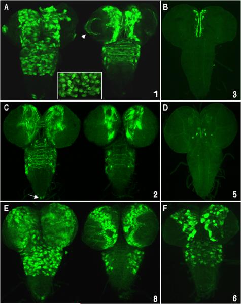
Fig. 6.
Many cas embryonic enhancers also direct gene expression during larval CNS development. CSC reporter Gal4 expression was detected using a Gal4 activated UAS/GFP-mCD8 transgene followed by anti-GFP fluorescent immunostaining. Images show dissected third-instar larval brains and ventral cords (anterior up) with panels A, C and E showing optical sections from the ventral (left) and dorsal (right) regions of the CNS. Panels B, D and F show stacked Z-series optical sections of the whole CNS. Numbers on the lower right side of each panel represent CSC regions shown in Fig. 2. A) cas-1 enhancer/reporter is expressed in many putative type II NBs and their lineages, including precursors and neurons of the central brain, thorax, and optic lobe neurons (arrowhead). cas-1 also directs expression in a cluster of abdominal ventral cord neurons located at the posterior tip (arrow). B) cas-3 enhancer/reporter expression in a subset of neurons within the medial brain hemispheres. Given their position and size, these medial-anterior neurons may be part of the neurosecretory cell group in the pars intercerebralis (de Valasco et al., 2007). C) cas-2 reporter is predominantly expressed in subsets of brain and thoracic neurons whose axons cross the midline, and in two neurons located at the posterior tip of the ventral cord (arrow). There are fewer GFP positive NBs compared with cas-1, suggesting that cas-2 activates expression predominantly in neurons. D) cas-5 enhancer/reporter activity was detected in 2 central brain neurons. E) cas-8 CSC activated reporter expression in a large subset of central brain lineages including optic lobe medullary NBs and their progeny, many thoracic lineages that project across the midline and posterior tip ventral cord neurons. F) cas-6 enhancer/reporter is expressed in a subset of Type II NB lineages.

