Figure 3.
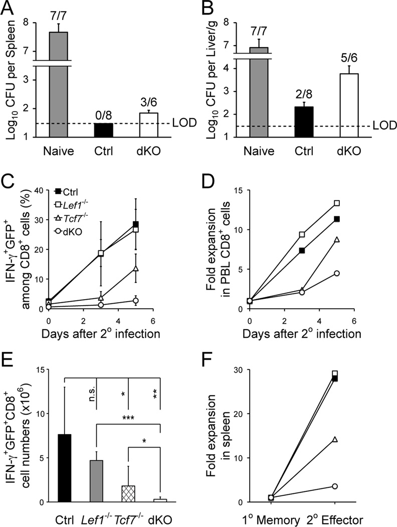
Loss of TCF-1 and LEF-1 greatly impaired recall response by the primary memory CD8+ T cells. A and B. Clearance of virulent LM-Ova by memory CD8+ T cells. Naïve or immune mice (day 35 post-infection) were challenged with virulent LM-Ova, and 3 days later, CFUs were determined in the liver and spleen. Shown are cumulative data from 2 experiments. Data are reported as CFU numbers (means ± s.d.) per spleen (A) or per gram of liver (B), from each organ with positive detection of LM-Ova. Frequency of animals with positive detection of the bacteria is marked on top of the bar. LOD, limit of detection. C and D. Secondary CD8+ T cell expansion in PBLs. Ova-specific CD8+ T cells were tracked in the PBLs after secondary challenge. (C) shows the frequency of IFN-γ+GFP+ cells among CD8+ T cells. (D) shows the relative expansion of secondary CD8+ effectors after normalization to starting memory CD8+ frequency. Data are representative of 3 independent experiments (n ≥ 7). E. Numbers of secondary effector CD8+ T cells in the spleens, as determined on day 5 after the re-challenge. Data are means ± s.d. from 3 independent experiments (n ≥ 5). *, p<0.05; **, p<0.01; ***, p<0.001; n.s., not significant. F. Relative expansion of secondary CD8+ effectors in the spleen. The numbers of secondary CD8+ effectors (as in E) was normalized to those of primary memory CD8+ T cells (as in Fig. 2A) to calculate the relative expansion.

