Figure 1.
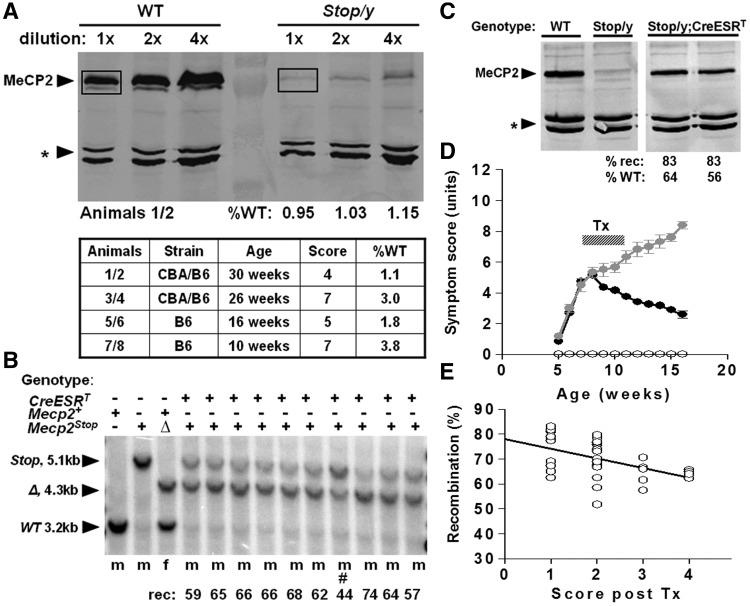
Silencing and rescue of MeCP2 expression in Mecp2Stop/y,Cre mice. (A) Quantitative western blots showing brain MeCP2 expression in wild-type (WT) and Stop/y males. Western blotting of whole brain extracts from wild-type and Stop/y littermates was used to quantify the amount of MeCP2 produced by the Stop allele. The amount of MeCP2 (boxed) was normalized to a non-specific cross-reacting band (asterisk) as a loading control. MeCP2 was measured for four Stop/y animals of different strain backgrounds, ages and aggregate symptom scores. (B) Southern blot showing recombination of the Stop/y allele (5.1 kb) to delete the Stop cassette (open triangle, 4.3 kb) after tamoxifen (Tx) treatment. All animals received the same tamoxifen-treatment regimen apart from #, which died soon after treatment started. (C) Representative western blot showing levels of MeCP2 protein in brains of wild-type, Stop/y and two tamoxifen-treated Stop/y;CreESRT animals. Recombination levels and the amount of protein relative to wild-type are shown. (D) Time plot showing phenotypic scores (see ‘Materials and methods’ section) for wild-type (open circle), Stop/y (black filled circles) and Stop/y,Cre mice (grey filled circles). Note the rapid symptom progression followed by a sustained improvement in symptom score following tamoxifen in the Stop/y,Cre cohort. (E) Recombination levels in brains of Stop/y,Cre mice showing relationship between higher recombination efficiency and milder symptom score at end of study.

