Figure 2.
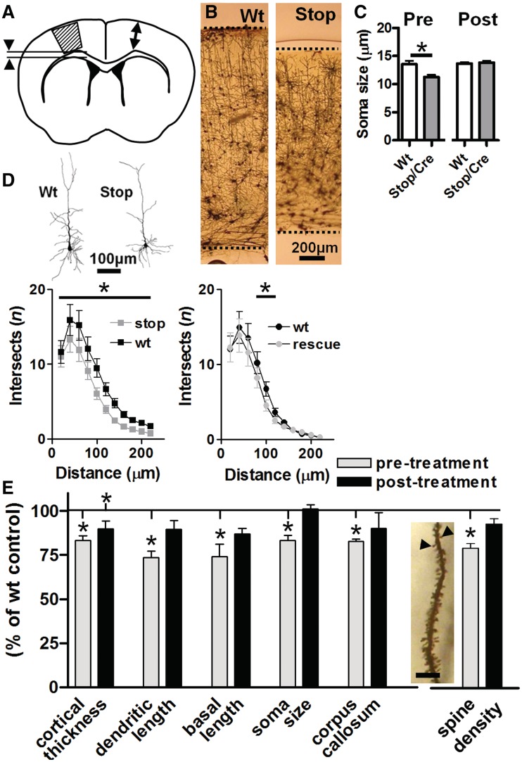
Quantitative assessment of neuronal morphology in primary motor cortex following Mecp2 reactivation. (A) Golgi labelled brain sections from wild-type (Wt) and Stop/y,Cre mice were sampled for gross measures of cortical thickness (arrows) and corpus callosum (arrowheads) as well as layer 2/3 pyramidal cell morphology within primary motor cortex (hatched area). Analysis revealed reduced cortical thickness (B, representative micrographs) and reduced soma diameter (C) in Stop/y,Cre untreated mice. (D) Quantitative assessment of reconstructed cells (representative examples shown) by Sholl analysis (n = 50–60 cells/four animals per group, total 210 cells) revealed reduced dendritic branching complexity in Stop/y,Cre mice compared with wild-type littermate controls at Sholl radii 20–140 µm from soma. At 8–10 weeks following tamoxifen treatment, overall branching complexity was still reduced (P < 0.05, ANOVA followed by Tukey A post hoc analysis) compared with wild-type but this difference was restricted to Sholl radii between 60–80 µm from soma. (E) Pooled data revealed a significant (t-test comparison with age-matched wild-type, *P < 0.05) reduction in all parameters measured (cortical thickness, total dendritic length, length of basal dendritic tree, soma diameter and corpus callosum thickness) in Stop/y,Cre mice compared with wild-type littermates. A reduction was also observed in spine density (measured in primary oblique dendrites of hippocampal CA1 pyramidal cells as shown in insert; scale = 10 μm). Analysis of tamoxifen treated (rescued) mice revealed no significant difference from their appropriate littermate controls with the exception of cortical thickness, which remained significantly reduced compared with age-matched controls.

