Figure 3.
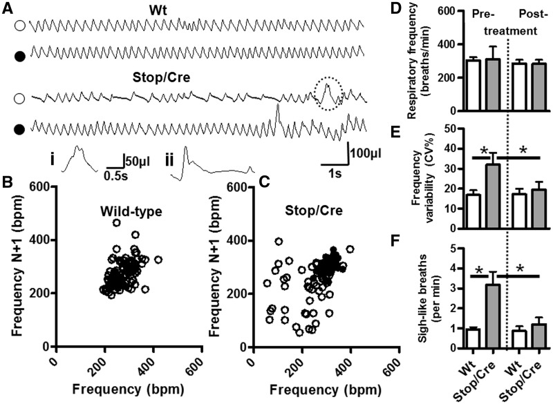
Improved respiratory pattern following delayed activation of Mecp2. (A) Representative plethysmographic traces of wild-type (Wt) and Stop/y,Cre mice before (open symbols) and 5 weeks following tamoxifen treatment (filled symbols). Pre-tamoxifen treatment, the respiratory trace from a Stop/y,Cre mouse is characterized by periods of fast to normal frequency breathing interspersed with periods of slow breathing due to prolonged expirations. Post-tamoxifen treatment, the respiratory trace from the Stop/y,Cre mouse is similar to wild-type. Insert (i) is an illustration of the sigh-like breath indicated by the dashed lines in the Stop/y,Cre breathing trace. Insert (ii) is an example of a typical sigh, large amplitude breath followed by a prolonged expiratory pause. These sigh-like breaths were highly prevalent in Stop/y,Cre respiratory traces. Plotting the respiratory traces as Poincare plots (B, wild-type; C, Stop/y,Cre) shows an increased scatter, thus increased variability of breathing pattern, in Stop/y,Cre pre-tamoxifen treatment (open symbols) when compared with post-tamoxifen Stop/y,Cre and with wild-type. Overall, there was no significant difference in average baseline (i.e. apnoea-free segments) respiratory frequency (D) between groups (wild-type, n = 11; Stop/y,Cre n = 8); however the variability in respiratory frequency (E) was significantly increased in Stop/y,Cre mice pre-tamoxifen treatment when compared with post-tamoxifen treatment and wild-type. Pre-tamoxifen treatment, Stop/y,Cre mice exhibited an increased number of sigh-like bpm [F, A(i) and A(ii)], which was significantly reduced post-tamoxifen treatment (*P < 0.05).

