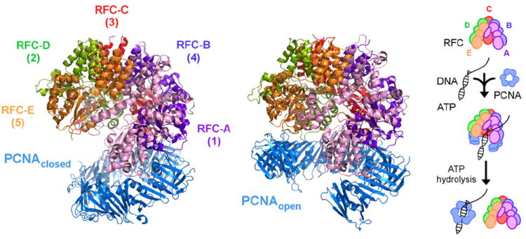
Fig. 1.

Model of the RFC·ATPγS·PCNA complex and PCNA loading reaction. Crystal structure of RFC bound to ATPγS and a closed PCNA clamp10 and a computationally derived model of RFC bound to ATPγS and open PCNA.42 The MD model represents current thinking about PCNA opening in a right-handed spiral conformation to allow entry of DNA during RFC-catalyzed clamp loading on ptDNA (minimal pathway depicted on the right). Note: RFC-A, RFC-B, RFC-C, RFC-D, and RFC-E correspond to RFC-1, RFC-4, RFC-3, RFC-2, and RFC-5, respectively.
