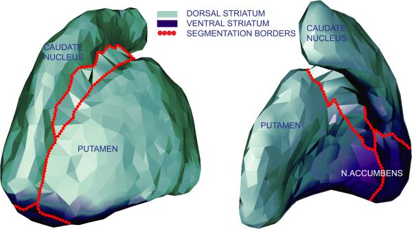
Figure 1. Anatomical and functional division of striatum.
Presented is a 3D model of the striatum, based on the segmentation of those voxels that were labeled striatum in > 20% of the segmentation masks. Model was created with GAMEs (Ferrarini et al., 2007) and displayed here for explanational purposes.

