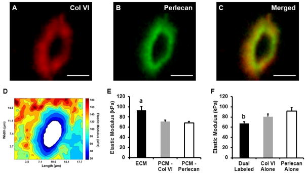
Figure 2.
AFM stiffness mapping of PCM regions dual-labeled for type VI collagen and perlecan. A representative PCM scan region in the middle/deep zone is shown. Dual IF-labeling for (A) type VI collagen and (B) perlecan demonstrated the (C) co-localization of these molecules in the pericellular space. Scale bar = 5 μm. (D) Contour map of calculated elastic moduli for the PCM scan region shown. (E) Elastic moduli of cartilage ECM and PCM as defined by the presence of type VI collagen or perlecan. There was no difference between biochemical definitions of the PCM (p = 0.70). ECM elastic moduli were significantly greater than PCM moduli (a: p < 0.005 as compared to either PCM definition). (F) Within the PCM regions, dual-labeled for type VI collagen and perlecan exhibited lower elastic moduli than regions positive for type VI collagen or perlecan alone (b: p < 0.05 as compared to type VI collagen and perlecan alone regions). Data presented as Mean + SEM (n ≥ 9).

