
Figure 2.
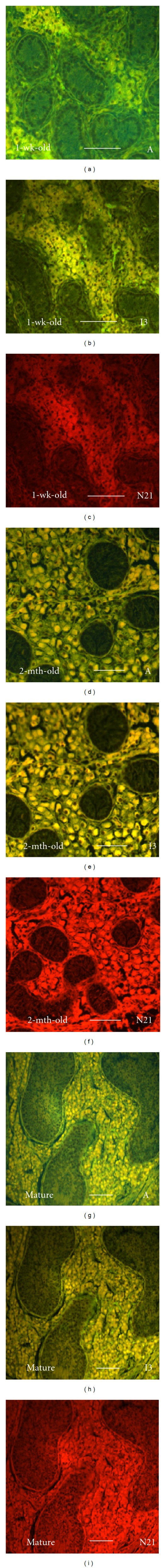
Autofluorescence in testis tissue sections from pigs of different ages examined using an epifluorescent microscope. Testis tissues from 1-wk-old (a–c), 2-month-old (d–f), and mature (g–i) pigs were fixed, sectioned, and examined for intrinsic fluorescence using an epifluorescent microscope equipped with filters of A (a, d, g), I3 (b, e, h), and N21 (c, f, i). Scale bars, 100 μm.