
Figure 7.
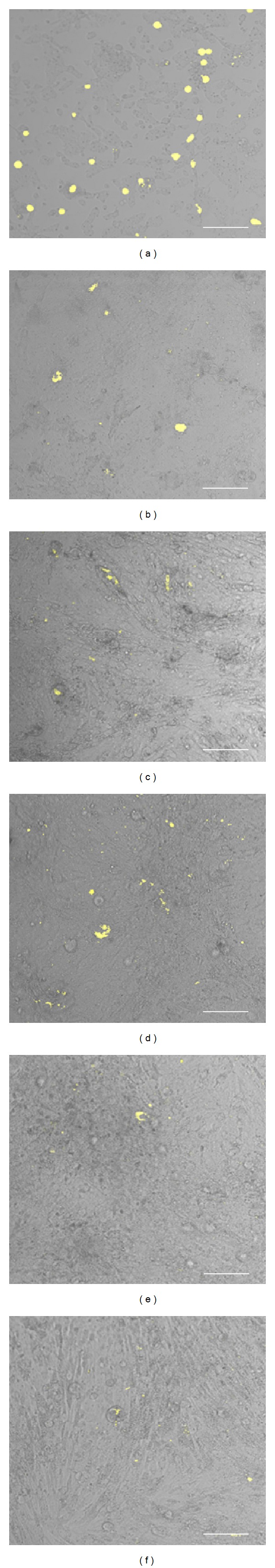
Autofluorescence in cultured testis cells. One-wk-old piglet testis cells were cultured in vitro for 6 days and examined for autofluorescence using a confocal laser scanning microscope and excited with a 405 nm laser and detection of emissions within 575–620 nm (yellow with brightfield overlay, (a–f) corresponding to 1–6 days following culture, resp.). Scale bars, 100 μm.