
Figure 9.
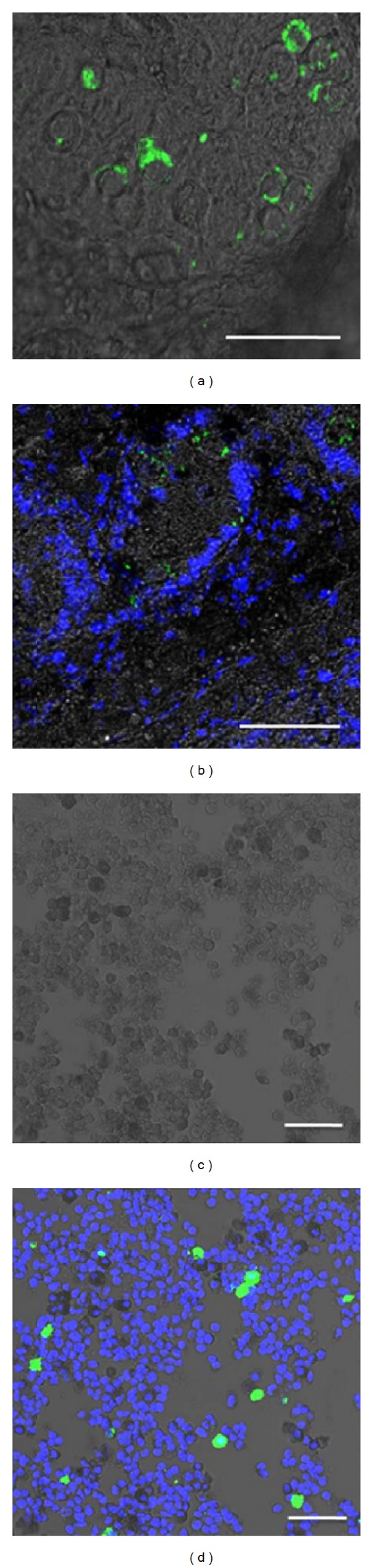
Identification of gonocytes with DBA staining, following the masking of autofluorescence by Sudan Black B in situ and in vitro. Piglet testis tissue sections (a, b) and dissociated cells (c, d) were stained with FITC-labeled lectin DBA and DAPI, followed by Sudan Black B staining and imaging with a confocal laser scanning microscope with brightfield overlay. Scale bars, 100 μm.