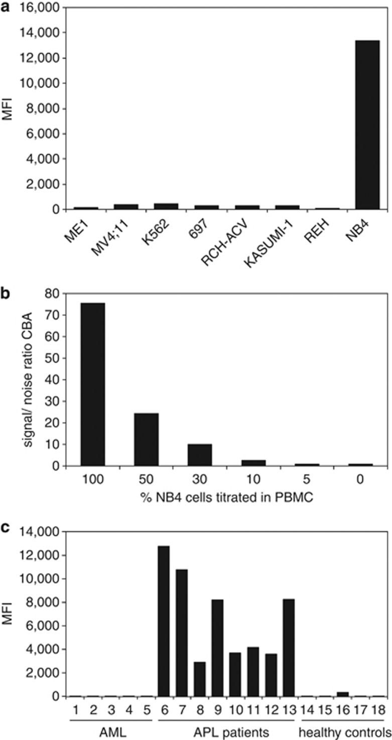
Figure 4.
Detection of PML–RARA fusion protein in cell lines and patient samples with the prototype PML–RARA immunobead assay. (a) Cell lysates from cell lines with different translocations expressing various fusion proteins: ME-1 with inv(16) expressing CBFβ-MYH11, MV4;11 with t(4;11) expressing MLL-AF4, K562 with t(9;22) expressing BCR-ABL, 697 with t(1;19) expressing E2A-PBX1, RCH-ACV with t(1;19) expressing E2A-PBX1, KASUMI-1 with t(8;21) expressing AML1-ETO, REH with t(12;21) expressing TEL-AML1 did not give a PE fluorescence signal in the PML–RARA immunobead assay, whereas the cell line NB4 with t(15;17) expressing PML–RARA (bcr1) gave a strong PE signal. (b) The PML–RARA positive NB4 cell line was diluted into PBMC of a healthy individual and cell lysates were prepared. The immunobead assay was carried out in triplicate for every diluted sample. At a concentration of 10% NB4 cells a signal-to-noise (s/n) ratio compared with the PBMC background of 2.7 was obtained. (c) The novel PML–RARA immunobead assay was carried out on a series of cell lysates of AML patients without the t(15;17) translocation (samples 1–5), APL patients with translocations in the PML gene involving both the bcr1 and bcr3 region and of two APL patients in which the break point region involved in the t(15;17) translocation was not determined (samples 6–13), and of healthy individuals (samples 14–19). Strong positive PE signals were only obtained in the cell lysates of APL patients independent of the bcr involved.

