
Fig. 4.
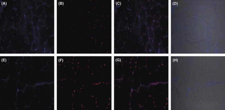
Transverse sections of quiescent gastrocnemius muscle of wild-type (A–D) and Sod1−/− (E–H) mice stained for p65 (blue, A and E), for nuclei with propidium iodide (red, B and F), an overlay of propidium iodide and p65 (C and G) and an overlay of light microscope image with p65 (D and H). Sections stained with secondary antibody alone were negative (data not shown).