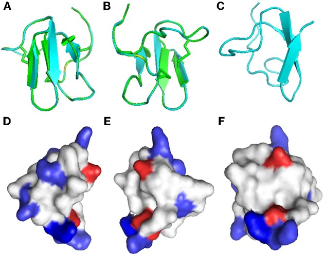
Figure 6.

Homology modeling of Juruin. Comparison of Juruin and U1-theraphotoxin-Ba1a (PDB ID: 2KGH), from Brachypelma ruhnaui, structures. (A,B) Juruin (blue) and U1-theraphotoxin-Ba1a (green) ribbon structures were superimposed over the backbone atoms. (C) Ribbon structure of Juruin in a different view related by ~90° rotation. (D,E) Molecular surface of Juruin highlighted to show electrostatic potential, surfaces with positive, negative, and neutral electrostatic potentials are drawn in blue, red, and white, respectively. (F) The Lys22-Lys23 segment shows maximum positive charge localization represented by the intense blue color. Model pairs show the sides of the protein rotated by ~180°.
