Figure 1.

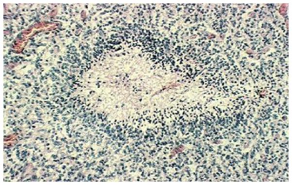
Hematoxyllin/eosin staining of human glioblastoma multiforme. Neoplastic cells stain blue and surround the area of central necrosis. Numerous microvessels are richly perfused with red blood cells.

Hematoxyllin/eosin staining of human glioblastoma multiforme. Neoplastic cells stain blue and surround the area of central necrosis. Numerous microvessels are richly perfused with red blood cells.