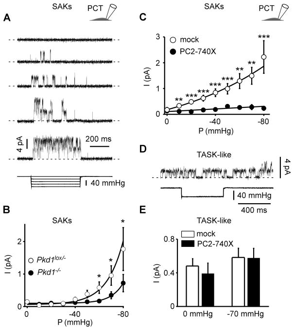
Figure 2. Polycystins regulate SAK activity in PCT cells.
A) Stretch-activated channels were recorded in the cell-attached patch configuration at a holding potential of 0 mV in immortalized mouse PCT cells. The pipette solution contained 5 mM K+ and the reversal potential of these channels was estimated to be about −80 mV (see Supp Fig. 1). Channel activity is illustrated at increasing negative pressure applied at the back of the patch pipette. Pressure pulses are shown at the bottom. Two channels were active in this patch. The zero current is indicated by a dashed line. B) Pressure-effect curves for mean pressure induced currents recorded in the cell-attached patch configuration in Pkd1lox/− (empty circles; n = 142) and in Pkd1−/− (filled circles; n = 146) PCT cells. Same conditions as in A. C) Mean SAK currents in PCT cells transiently transfected with either a mock empty pIRES2-DsRed plasmid (empty circles; n = 97) or together with PC2-740X (filled circles; n = 82). Currents were recorded in the cell-attached patch configuration at a holding potential of 0 mV. D) TASK-like channel activity recorded in the same conditions as in A. These channels are not responsive to an increase in pressure as shown in the lower trace. E) The histogram shows the effect of PC2-740X (n = 31) as compared to the mock empty expression vector (n = 33) on TASK-like channel activity in PCT cells. Data represent mean ± standard error of the mean.

