
Fig. 1.
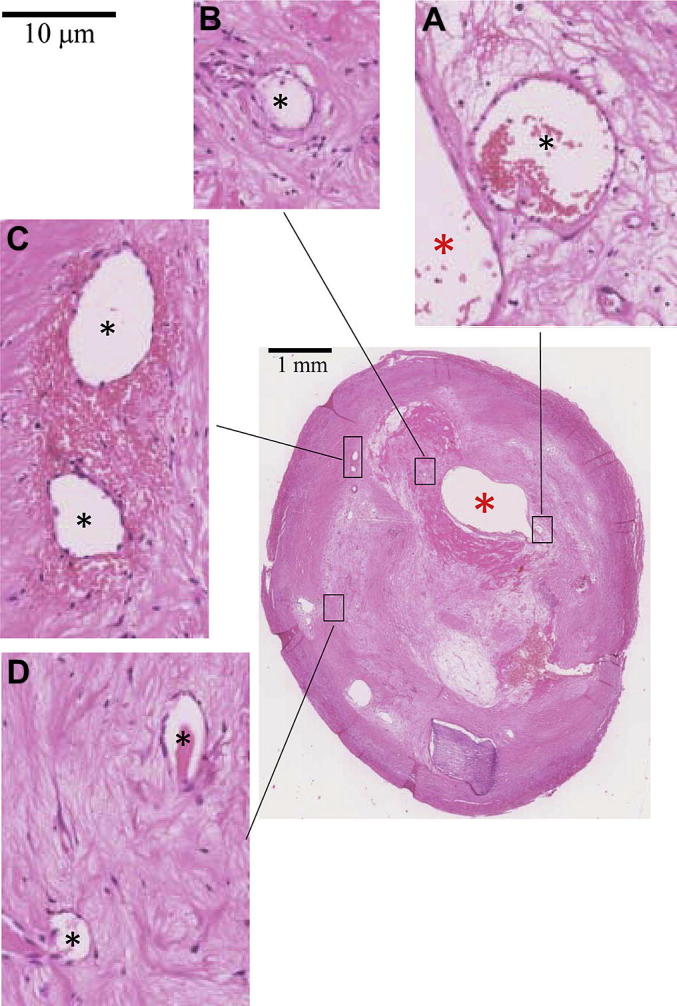
Microscopic slide (H&E) showing plaque structures (A&B: neovessels closed to the main arterial lumen; C: neovessels located in the middle region with abundant adjacent red blood cells; and D: neovessels located in a peripheral region; red asterisk stands for the main arterial lumen and black asterisk for the lumen of neovessel).