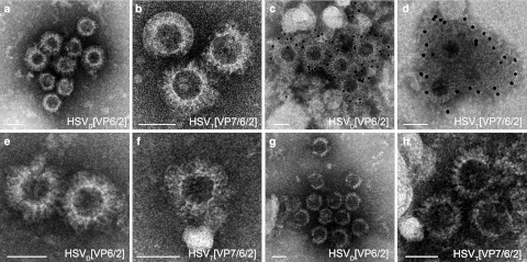
Figure 4.
Electron micrographs of herpes simplex virus type 1 (HSV-1) amplicon vector encoded rotavirus-like particles (RVLPs). Purified RVLPs from (a–d) Vero 2-2 cells, (e,f) HeLa cells, and (g,h) Hek293 cells infected with HSV-1 amplicon vectors (MOI 1). Two days postinfection, RVLPs were purified over a sucrose cushion and the concentrated particles were analyzed by electron microscopy. (a,b) Negative staining of RVLPs from Vero 2-2 cells infected with (a) HSVD[VP6/2] and (b) HSVT[VP76/2]. (c,d) Immunogold staining using a polyclonal anti-RV serum and a secondary antibody coupled to 12-nm gold particles. (c) Double-layered RVLPs from cells infected with HSVD[VP6/2]. (d) RVLPs from cells infected with HSVT[VP7/6/2]. (e,f) Negative staining of RVLPs from HeLa cells infected with (e) HSVD[VP6/2] and (f) HSVT[VP76/2]. (g,h) Negative staining of RVLPs from Hek293 cells infected with (g) HSVD[VP6/2] and (h) HSVT[VP76/2]. Bars = 50 nm.

