Figure 5.
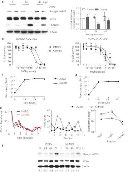
L4-100K promotes viral mRNA translation. (a) ID8 CuO-100K cells were infected with dl922-947 (MOI 10) in the presence or absence of cumate (200 µg/ml) for up to 48 hours. Expression of eIF4E, phospho-eIF4E, and L4-100K was assessed by immunoblot. Relative eIF4E phosphorylation in two separate blots was quantified using ImageJ (right). (b) ID8 CuO-100K and CMT64 CuO-100K were infected with dl922-947 (MOI 0.01–10,000) in the presence and absence of cumate. Cell survival was assessed 120 hours postinfection by MTT assay. (c,d) ID8 CuO 100K were infected with dl922-947 (MOI 10) in the presence and absence of cumate. (c) Viral genomes and (d) Hexon transcripts were quantified using quantitative PCR up to 72 hours postinfection. (e) ID8 CuO-100K cells were infected with dl922-947 (MOI 10) in the presence of DMSO or cumate. Forty-eight hours later, ribosomal fractions were prepared as before (left). RNA concentration was quantified in each fraction (center). Hexon transcript number in pooled fractions was quantified using qRT-PCR (right). *P < 0.05. (f) ID8 CuO-100K cells were infected with dl922-947 (MOI 10) in the presence of DMSO or cumate for up to 72 hours. Expression of eIF2α and phospho-eIF2α was assessed by immunoblot. Extent of phosphorylation was assessed using ImageJ—numbers below each lane represent phospho-eIF2α:total eiF2α ratio normalized to uninfected cells. DMSO, dimethyl sulfoxide; h.p.i., hours postinfection; MOI, multiplicity of infection; MTT, 3-(4,5-dimethylthiazol-2-yl)-2,5-diphenyl tetrazolium bromide; pfu, plaque-forming unit; qRT-PCR, quantitative reverse transcription-PCR.

