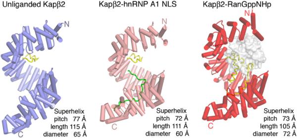
Figure 1. Ribbon Diagrams of Unliganded and Substrate- and Ran-Bound Kapβ2s.
β helices are represented as cylinders and structurally disordered loops as dashed lines. Unliganded Kapβ2 is in blue, Kapβ2 bound to substrate is in pink, Kapβ2 bound to Ran is in red, and H8 loops in all three structures are in yellow. Substrate hnRNP A1-NLS is in green and Ran is drawn as a surface representation in gray.

