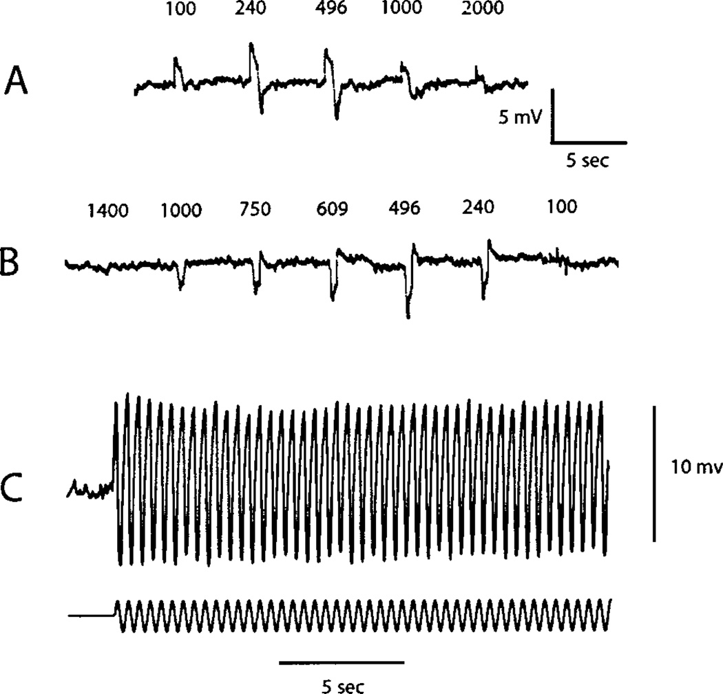
Fig. 1.
Response of an ON bipolar cell to a contrast step of variable diameter (A) and variable inner diameter of a concentric annulus (B). Dimensions in microns are shown above each response. The 100-µm spot and all other stimuli are centered in the receptive field. The contrast step is 0.3 log unit, that is, two times greater than the steady background of 20 cd/m2. (C) Response to a sinusoidal modulation of 80% at a frequency of 3 Hz and diameter of 240 µm.

