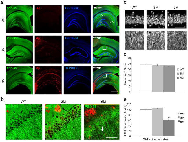
Figure 1. PSD-95 decreases in apical dendrites in 6 month old, but not in 3 month old, 5XFAD mice.
a. PSD-95 (green) and Aβ (red) immunoreactivity with TO-PRO 3 (blue) nuclear counterstain in hippocampus of wild type (WT), 3 month old (3M) and 6 month old (6M) 5XFAD mice. PSD-95 immunoreactivity highlights regions of apical dendrites in CA1, CA3 and DG (dentate gyrus). Boxes indicate the portion of CA1 region illustrated in b and c. Bar for all = 500 μm. b. High power images show PSD-95 and Aβ double labeling. PSD-95 immunoreactivity is prominent in apical dendrites and weak in soma in WT. Similar PSD-95 distribution is seen in 3M 5XFAD, despite of the presence of Aβ plaques. In contrast, PSD-95 immunoreactivity is weak in apical dendrites and prominent in soma (arrow) in 6M 5XFAD. Bar for all = 50 μm. c. Images of 100 μm × 100 μm areas show TO-PRO 3 stained nuclei (upper panel) and PSD-95 labeled apical dendrites (lower panel) that were analyzed in d and e respectively. Bar for all = 50 μm. d. Numbers of neuronal nuclei counted in CA1 pyramidal layer do not differ among the 3 groups: WT = 24.0±0.4; 3M = 23.5 ±0.5; 6M = 23.0±0.6 (p > 0.05, ANOVA). e. PSD-95 intensity measured in CA1 apical dendrites is reduced in 6M 5XFAD by 39.6±1.3% and 41.8±1.4% compared with WT and 3M 5XFAD mice, respectively (*: p < 0.001, post hoc Tukey test).

