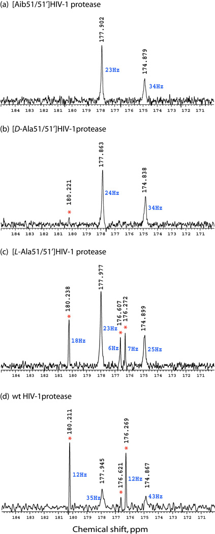
Fig. 3.
226 MHz 13C-{1H} NMR spectra for unliganded HIV-1 protease and three chemically synthesized analogue enzymes. (a) [Aib51/51’]HIV-1 protease, (b) [D-Ala51/51’]HIV-1 protease, (c) [L-Ala51/51’]HIV-1 protease, (d) wild-type HIV-1 protease. Red asterisk indicates peaks originating from peptide autoproteolysis products. Linewidths of the peaks are in blue. All samples were prepared in 18.9 mM Na.phosphate buffer (pH 5.7), containing 5.4 % (v/v) D2O and 100 µM DSS-d6. Concentrations of protein were 0.29 – 0.41 mM.

