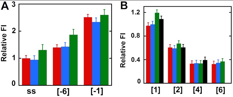
Fig. 3.
Fluorescence intensity changes caused by helicase binding for forked DNA constructs with 2-AP monomer probes in the leading (3′ → 5′) strand. (A) Intensities at 370 nm for ssDNA and for constructs with 2-AP monomer probes located directly at the ss–dsDNA fork junction. (B) Intensities for 2-AP monomer probes located deeper within the dsDNA portion of the construct. Color coding is the same as in Fig. 2.

