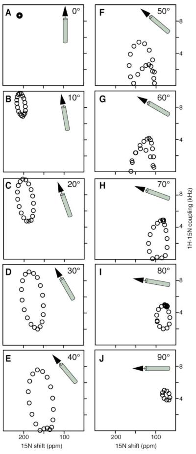
FIG. 1.
PISEMA spectra calculated for a 19-residue α-helix with 3.6 residues per turn and uniform dihedral angles at various helix tilt angles relative to the bilayer normal. A. 0°. B. 10°. C. 20°. D. 30°. E. 40°. F. 50°. G. 60°. H. 70°. I. 80°. J. 90°. Spectra were calculated on a Silicon Graphics O2 computer (Mountain View, CA), using the FORTRAN program FINGERPRINT (21, 24). The principal values and molecular orientation of the 15N chemical shift tensor (σ11 = 64 ppm; σ22 = 77 ppm; σ33 = 217 ppm; σ33NH = 17°) and the NH bond distance (1.07 Å) were as previously determined (19).

