Figure 1.
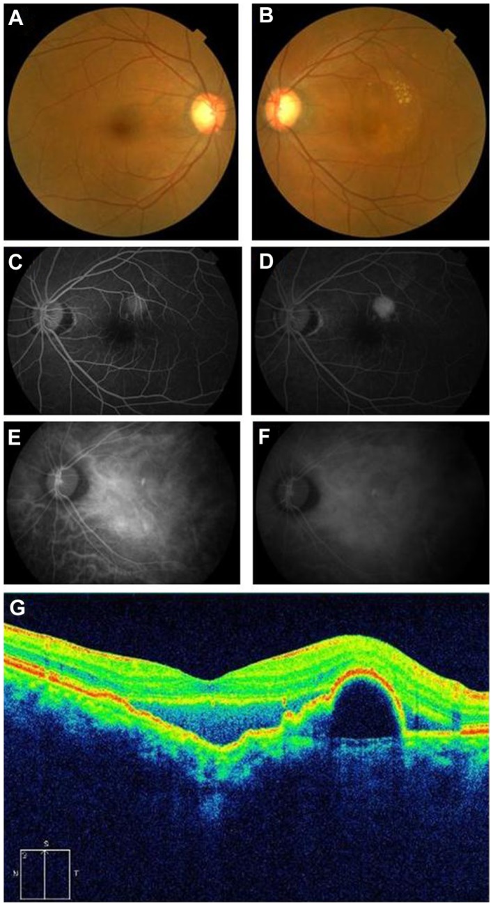
(A and B) show color fundus photographs of the right and left eyes, respectively. The left eye shows serous retinal detachment and exudates. (C and D) show the results of FA (early and late phase, respectively) and show hyperfluorescence (window defect) at the lesion of the PED. (E and F) show the results of IA (early and late phase, respectively) and show polypoidal fluorescence at the yellowish protruding lesion. SD-OCT demonstrates the separation of the retina between the inner segment and the outer segment junctions of the photoreceptor (IS/OS) line and the RPE with choroidal excavation (G).
Note: A protruding lesion was observed in the upper macular area, and PED ran along the upper part.
Abbreviations: FA, fluorescein angiography; PED, pigment epithelium detachment; SD-OCT, spectral-domain optical coherence tomography; IS/OS, inner segment/outer segment; IA, indocyanine green augiography; RPE, retinal pigment epithelium.

