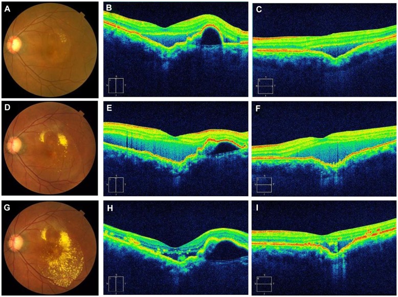
Figure 2.
(A–C) are fundus photographs and OCT images before anti-VEGF treatment. (D–F) are fundus photographs and OCT images after initial anti-VEGF treatment. (G–I) are fundus photographs and OCT images after three rounds of anti-VEGF treatment. (B, E and H) are vertical OCT images and (C, F and I) are horizontal OCT images. The left eye shows serous retinal detachment and exudates (A). A choroidal excavation is located just inside the macular area. As described in Figure 1, the OCT images show the separation of the retina between the IS/OS line and the RPE (B). The horizontal image shows choroidal excavation more clearly than the vertical image (C). The left eye shows more exudates than that of her initial visit to us (A) (D) and an increase in serous retinal detachment (E and F). The retinal exudates increased after the 3 injections (G), but serous retinal detachment decreased (H and I).
Note: The extent of PED may have increased.
Abbreviations: OCT, optical coherence tomography; VEGF, vascular endothelial growth factor; IS/OS, inner segment/outer segment; RPE, retinal pigment epithelium; PED, pigment epithelium detachment.

