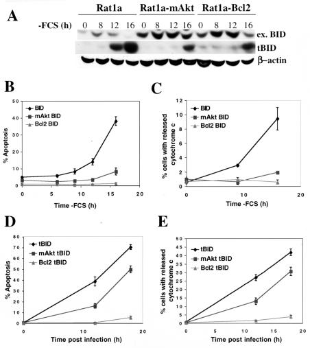
FIG. 2.
Activated Akt, like Bcl-2, inhibits mitochondrial cytochrome c release and apoptosis following BID cleavage. (A) Rat1a, Rat1a-mAkt, and Rat1a-Bcl-2 fibroblasts stably overexpressing BID were deprived of serum, and BID cleavage was monitored by immunoblotting using anti-BID and anti-tBID antibodies. ex. BID, exogenous BID. (B) Kinetics of apoptosis following serum deprivation in cell lines overexpressing BID. (C) Kinetics of cytochrome c release following serum deprivation in cell lines overexpressing BID. The experiment was performed as in panel B, except that cells were fixed and immunofluorescence for cytochrome c was performed. Cells exhibiting an extramitochondrial, diffuse pattern of cytochrome c staining were scored positive for cytochrome c release as described in Materials and Methods. (D) Rat1a, Rat1a-mAkt, and Rat1a-Bcl-2 cell lines were infected with tBID-expressing retrovirus, as described in Materials and Methods, and apoptosis was quantitated, as for panel B, 12 and 18 h postinfection. Apoptosis was quantitated as for panel B. (E) Cytochrome c was quantitated following infection with retrovirus expressing tBID. The experiment was performed as for panel C, except cells were infected in the presence of 100 μM caspase inhibitor zVAD. Cells were fixed and immunofluorescence for cytochrome c was performed. Cells exhibiting an extramitochondrial, diffuse pattern of cytochrome c staining were scored positive for cytochrome c release. All data are depicted graphically as the means ± standard errors of the means for three independent experiments.

