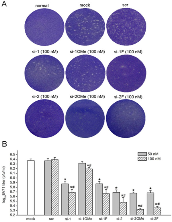
Figure 6.
Determination of virus titers by plaque assays. RD cells were transfected with unmodified and 2′-modified siRNAs targeting the 5′ UTR of the EV71 genome and then infected with 0.01 MOI of EV71. At 48 h post-infection, culture cell supernatants were collected. Monolayers of RD cells in 6-well plates were inoculated with 500 μl of supernatants at a 10-4 dilution for 1 h, and then plaque assays were carried out to determine virus titers. Culture supernatants from non-infected normal cells and mock transfection cells were used as negative and positive control, respectively. (A) Representative plaque formation on RD cell monolayers. (B) Virus titers in culture supernatants. Values are means ± SD of three independent experiments. *P < 0.05, compared with mock transfection control. #P < 0.05, compared with 50 nM of si-1, si-1OMe, si-1 F, si-2, si-2OMe and si-2 F, respectively.

