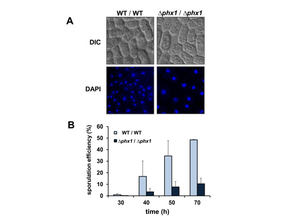
Figure 6.
Sporulation defect of Δphx1/Δphx1 mutant diploid. (A) The wild type and mutant diploid cells were grown to the stationary phase (OD600 of 8–9; ~70 h culture) in EMM at 30°C and examined under the microscope (Axiovert 200 M, Carl Zeiss). Representative DIC and DAPI images were presented. (B) Quantification of the sporulation efficiency. Diploid cells grown for different lengths of time at 30°C in EMM were examined under the microscope to count the number of spore-containing asci. The percentage of asci formation among a total of more than 500 counted cells was presented as sporulation efficiency. Cells grown from three independent cultures were examined to obtain average values.

