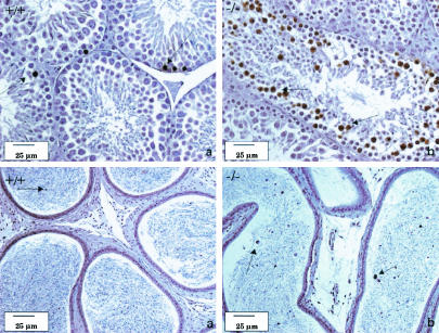
FIG. 4.
Formalin-fixed sections of the testes (top) and epididymides (bottom) of LGR7 males (tissues stained with the TUNEL method). (a) Wild-type male. Shown is normal regular apoptosis of spermatogenic cells in the seminiferous tubules of the testis (arrows); some apoptotic spermatogenic cells in the acini of the epididymis (arrow) are also shown. (b) LGR7−/− male. Shown are apoptotic stage 12 meiotic cells in the testes (arrows) and increased numbers of apoptotic cells in the lumina of the epididymis (arrows).

