
Figure 4.
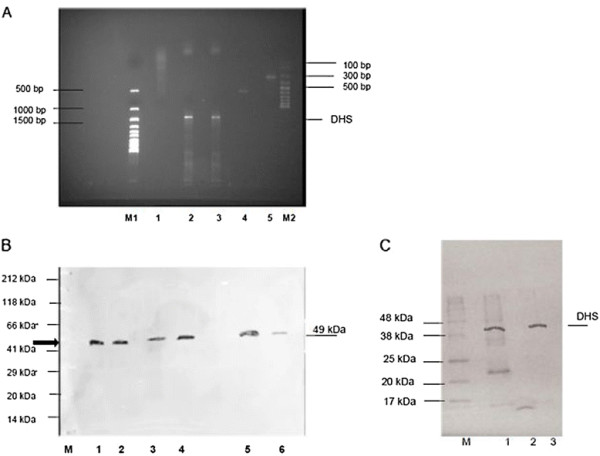
A) Monitoring the in vivo knockdown ofP. bergheiinfected schizonts transgenic for the expressed plasmodial DHS shRNA by RT-PCR two days post infection with NMRI mice. NMRI mice were infected with transgenic schizonts transfected with the plasmodial shRNA P#176 construct. M1) 1 kb ladder (LifeTechnologies, Karlsruhe, Germany); 1) DHS-shRNA; 2) EIF-5A-shRNA; 3) Amplification of the recombinant pcDNA3 vector carrying the dhs gene from P. falciparum generates a cDNA fragment of 1491 bp. 4) Quality control of total, cellular RNA by amplification of a 548 bp fragment with α-tubulin gene-specific primers from P. berghei; 5) PCR-control of recombinant eIF-5A (448 bp) expression vector with eIF-5A primers; M2) 100 bp ladder (LifeTechnologies, Karlsruhe, Germany) B)In vivo silencing of plasmodial DHS monitored by Western blot analysis after infection of NMRI mice with transgenic schizonts expressing shDHS. 1 and 2) Two different concentrations of purified, human DHS protein; 3) PB ANKA wild type strain protein extract 4) Mock strain protein extract; 5) eIF-5A shRNA P#18; 6) DHS- shRNA P#176. C) Validation of anti-DHS antibody specificity in crude protein extracts prepared after infection of NMRI mice expressing plasmodial DHS #176 shRNA construct. M) Blueeye Marker; 1) crude protein extract from infected NMRI mice with plasmodial DHS #176 shRNA construct and supplemented with recombinant human protein; 2) crude protein extract from infected NMRI mice with plasmodial DHS #176 shRNA construct; 3) purified recombinant human DHS protein. The protein concentration was 10 μg in each lane.