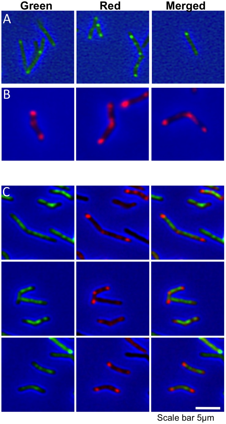
Figure 2. Co-localization of PBP1a and VanBODIPY in M. smegmatis.
(A) Three fields of uninduced M. smegmatis mc2155 pMEND-PBP1a-mCherry stained with VanBODIPY display the characteristic polar and septal staining of nascent peptidoglycan (Green spots). (B) Induction of PBP1a-mCherry with 20 ng/ml Tc for 3.5 hr results in strong expression of red PBP1a-mCherry that localizes to the septum and poles, in a pattern similar to VanBODIPY staining (three fields of cells). (C) Expression of PBP1a-mCherry disrupts the localization of VanBODIPY staining, leaving diffuse green staining across the cell. The columns show the green, red and merged images respectively.

