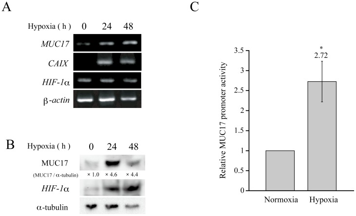
Figure 1. MUC17 expression is enhanced by hypoxia.
(A) AsPC1 cells were cultured under hypoxic conditions (1% O2) for the indicated times. MUC17 mRNA expression was examined by RT-PCR at each time point. (B) AsPC1 cells were cultured under normoxic or hypoxic conditions for the indicated times. Cell lysates were probed with anti-MUC17, HIF1α, and α-tubulin antibodies by Western blot analysis. The intensities of the bands were quantitated by densitometric scanning, and the ratio of MUC17 to α-tubulin expression is shown under each band as the relative intensity compared with that obtained in normoxic AsPC1 cells. (C) MUC17 promoter activity was measured by a Dual-Luciferase Reporter Assay. After transfection of the MUC17 reporter plasmid, AsPC1 cells were incubated under normoxic or hypoxic conditions for 24 h. Cell lysates were assayed using a luciferase assay kit in a Tristar multimode microplate reader LB941 (Berthold Technologies). Transformation efficiency was normalized on the basis of Renilla luciferase activity. The promoter activity under normoxic conditions was given a value of 1. P values were determined using the Student's t-test. * P<0.05.

