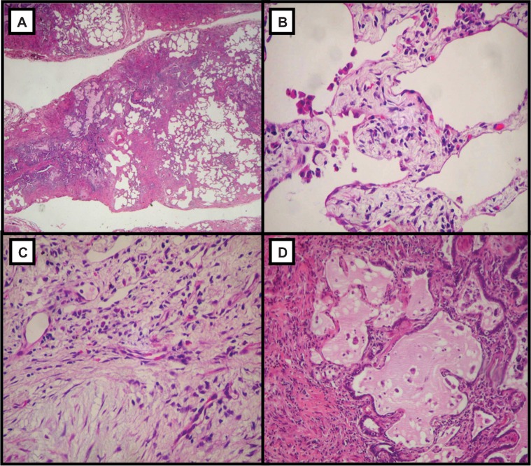
Figure 1.
Panoramic view showing unaffected areas alternating with fibrous and cystic changes, which characterize the UIP histological pattern (A, ×40); collapsed area with alveolar collapse and interstitial mononuclear infiltrates (B, ×100); mural fibrosis area with septal fibromyxoid tissue and fibroblastic foci (C, ×100); and honeycombing area with irregular cystic air spaces between bands of fibrous connective tissue (D, ×100). Hematoxylin and eosin staining.

