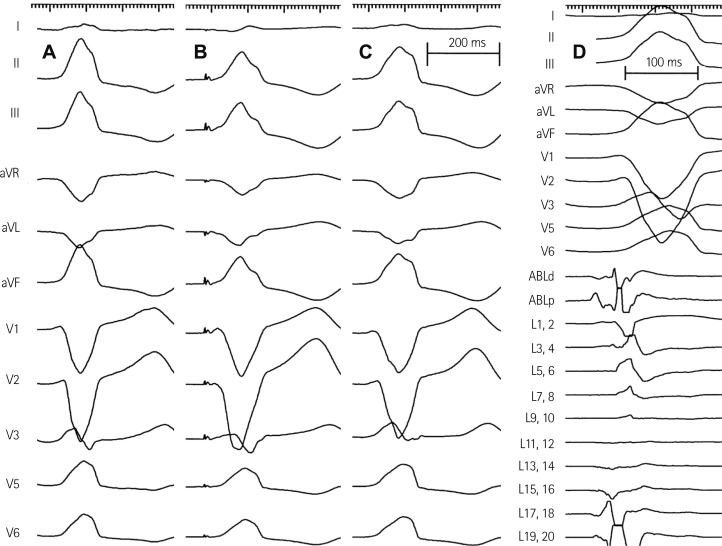
Fig. 3.
Activation and paced mapping for radiofrequency catheter ablation. A: spontaneous PVCs during electrophysiologic study had left bundle branch block configuration and inferior axis. B: paced mapping at anterolateral wall of RVOT 2 cm below pulmonary valve showed the QRS morphology nearly identical with spontaneous PVCs. C: the morphology of accelerated ventricular rhythm induced by radiofrequency ablation also had the QRS morphology nearly identical with spontaneous PVCs. D: activation mapping for spontaneous PVCs showed the earliest ventricular activation signal 20 ms prior to the QRS onset. PVC: premature ventricular complex, RVOT: right ventricular outflow tract, ABLd: distal ablation catheter, ABLp: proximal ablation catheter, L: Lasso variable circular mapping catheter.

