Fig. 1.
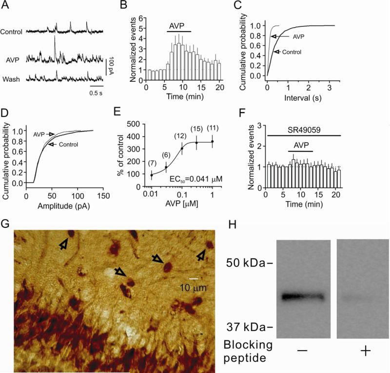
AVP increases the frequency not the amplitude of sIPSCs recorded from the CA1 pyramidal neurons of the hippocampus via activation of V1A receptors. A, sIPSCs recorded from a CA1 pyramidal neuron before, during and after the application of AVP (0.3 μM). B, Time course of the sIPSC frequency averaged from 9 cells. C, Cumulative frequency distribution from a CA1 pyramidal neuron before and during the application of AVP. D, Cumulative amplitude distribution from the same cell before and during the application of AVP. E, Concentration-response curve of AVP. Numbers in the parenthesis are numbers of cells recorded. F, Pretreatment of slices with and continuous bath application of the selective V1A receptor antagonist, SR49059 (1 μM), blocked AVP-induced increases in sIPSC frequency (n=8). G, Immunoreactivity of V1A receptors was detected in both pyramidal neurons and interneurons in CA1 region. Arrows indicate interneurons in the stratum radiatum. H, Western blot demonstrated the expression of V1A receptors in the lysates of the CA1 region. A band of ~43 kDa was detected whereas pretreatment of the V1A antibody with the corresponding blocking peptide significantly reduced the density of the band demonstrating the specificity of the antibody.

