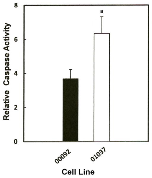
Fig. 6.

Effect of Mn on caspase-3 activity in lymphocyte cell lines, ND00092 and ND01037. Cells were treated with the 0.5 mM Mn for 12 hrs. Fifty μl of the cell lysate from each sample was added to the 96 well plates in triplicate and incubated with Z-DEVD-AMC for 30 minutes at room temperature. All activities measured were normalized to protein concentration and fluorescence was measured using the CytofluorII Multiplate Reader with excitation set at 380 nm and emission set at 460 nm. Data is the mean ± S.E of three independent experiments. Results indicate there was significant increase in the relative caspase activity in ND01037 when compared to ND00092, ap < 0.025.
