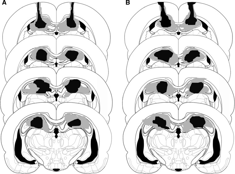
Fig. 2.
An example lesioned area in dorsal hippocampus verified by histology (the arrowhead). A lesion area before extinction in experiment 2. B lesion area just after extinction in experiment 3. Minimum (black) and maximum (gray) extent of dosal hippocampus lesions in animals Experiments. The lesions were reconstructed on successive coronal sections (−2.4, −3.0, −3.6, and −4.2 mm to bregma) from Paxinos and Watson (2005)

