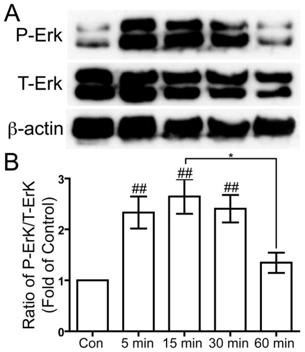
Figure 5. US-induced phosphorylation of Erk at threonine 202 and tyrosine 204.
Serum-deprived primary human chondrocytes were treated with US for three minutes, then total cell lysates were collected 5, 15, 30 and 60 minutes after US treatment. Total cell lysate for control was collected from chondrocytes that did not receive US treatment. Phosphorylation of Erk1/2 at threonine 202 and tyrosine 204, total Erk1/2 and β-actin loading control were demonstrated by Western blotting (A). Western blotting data from six separate experiments were quantified using Image J and averages are shown as the ratio of phosphorylated to total Erk (B). * p < 0.05. ## p < 0.01 vs. control.

