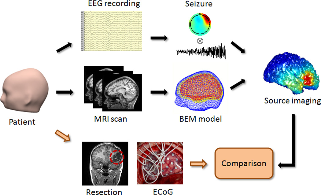
Fig. 1.

Schematic diagram of seizure source imaging and the study design. Seizure activity from scalp EEG recordings was analyzed to image seizure sources. Patient-specific boundary element head models were created from the pre-operative MRI images of the patients. Surgical resections and intracranial recordings were used to validate the seizure source imaging results.
