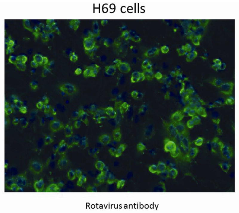
Figure 2. Immunohistochemistry for RRV infection of H69 cells.

Staining with anti-rotavirus antibody, shown in green, demonstrates the presence of live RRV in the immortalized human cholangiocytes 24 hours post inoculation. Cell nuclei are counter stained with DAPI, shown in blue. Magnification 20X.
