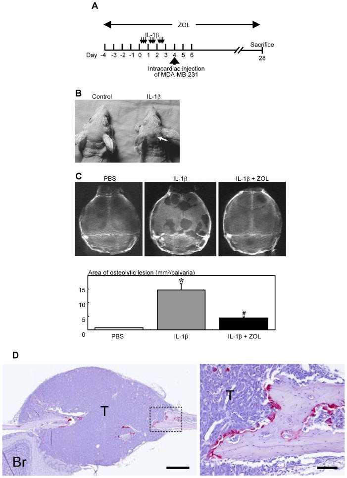
FIGURE 1. Effects of bone resorption on the development of bone metastases of MDA-MB-231 human breast cancer cells in nude mice.
(A) Experimental protocol. Four-week-old female athymic nude mice received repeated subcutaneous injections of IL-1β (thin arrows, 2 μg/10 μl/injection) over the calvariae 3 times a day for 3 days. Twenty-four hours after the last injection, MDA-MB-231 cells were inoculated into the left cardiac ventricle (thick arrow). In one group of animals ZOL (4 μg/mouse, s. c., daily) was given from 4 days before the IL-1β injection to the end of the experiments. Mice were sacrificed at day 28.
(B) Macroscopic view of tumor formation at the time of sacrifice. Note tumor formation on the head of IL-1β-treated mouse (arrow).
(C) Representative radiographic view of calvariae and quantitative analysis of osteolytic lesions in PBS-, IL-1β-, and IL-1β + ZOL-treated mice. Data are mean from two separate experiments (n = 6 mice x 2 experiments = 12 for each group).
* Significantly greater than PBS group (p < 0.01).
# Significantly less than IL-1β group (p < 0.01).
(D) Left: Representative histological view of metastasis in calvariae. Right: Higher magnification of the dashed rectangle area. Tumor colonization (T) is associated with calvarial bone destruction with numerous TRAP-positive osteoclasts along the surface of adjacent bone. (TRAP staining, Br: brain, scale bars = 500 μm (left), 100 μm (right)).

