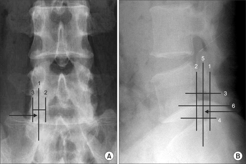
Fig. 2.
(A) The anterior-posterior view of the lumbar spine, with the superimposed line (1) bisecting the pedicle. This line was drawn halfway between the farthest medial (2) and the farthest lateral (3) points on the pedicle. (B) The lateral view of the lumbar spine with the quadrant system superimposed. A line was drawn tangent to the curve of the spine at the level of interest along the posterior vertebral line. (1) A second line (2) was drawn parallel to the third line at the posterior margin of the foramen. Next, two lines perpendicular to lines 1 and 2 were drawn at the superior and inferior margins of the foramen (lines 3 and 4, respectively). Finally, line 5 was drawn bisecting lines 1 and 2, and line 6 was drawn bisecting lines 3 and 4. These lines divided the foramen into four quadrants. Arrow: needle position.

