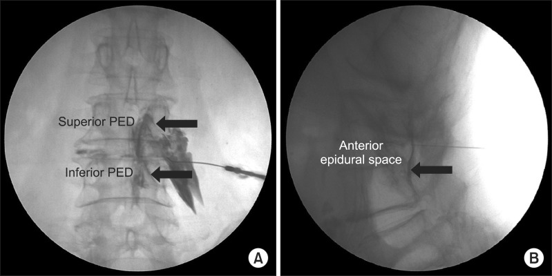
Fig. 3.
(A) The anteroposterior view of a needle placed for a Kambin's triangle approach of the L5 nerve root demonstrating the landmarks used for this investigation. Note the contrast flowing along the most medial aspect of the inferior pedicle (PED). (B) The lateral view of a needle placed for a Kambin's triangle approach of the L5 nerve root demonstrating the landmarks used for this investigation. Note the contrast flowing anterior epidural space.

