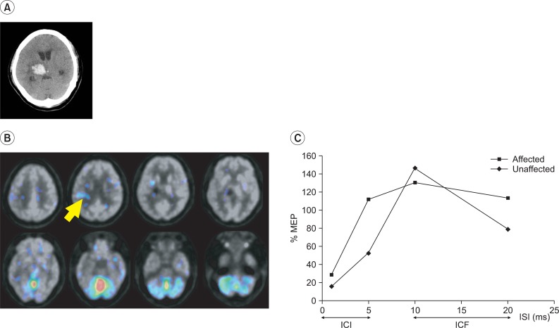
Fig. 1.
A patient in a good recovery group (Patient 4). (A) Brain CT showed a hemorrhagic lesion in the right thalamus. (B) During the motor task, activation was observed in the cerebellum and perilesional motor cortex (arrow) on her FDG-PET. (C) Normal ICI and ICF response patterns were observed in bilateral motor cortices on the paired pulse TMS. ICI: Intracortical inhibition, ICF: Intracortical facilitation, ISI: Interstimulus interval, ms: Milliseconds.

